How exercise affects your eyes

Regular exercise has long been recognized as a way to improve overall health and well-being. Exercise can reduce the risk of heart disease, stroke and obesity, among other ailments. However, what many people do not realise is that exercise can also have a positive impact on our eyes.

Studies have shown that regular exercise can reduce the risk of developing serious eye diseases that may lead to blindness. These diseases include diabetic retinopathy, glaucoma and macular degeneration, which are often linked to high cholesterol level, diabetes and high blood pressure. By reducing these risk factors, exercise can help preserve vision for a longer period.

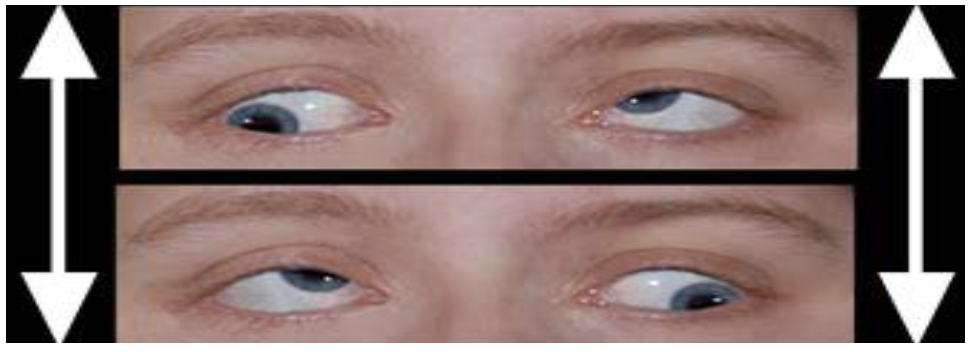
While no direct correlation has been found between exercise and glaucoma, research has shown that intraocular pressure is reduced after exercising. This is due to the acceleration of aqueous outflow, leading to lower intraocular pressure than usual. People who regularly exercise have a lower risk of developing glaucoma than those who do not. According to the American Academy of Ophthalmology, people who engage in moderate physical exercise are 25% less likely to develop glaucoma than those who are mostly inactive. However, caution should be exercised as certain positions and postures can exacerbate glaucoma, such as a head-stand.

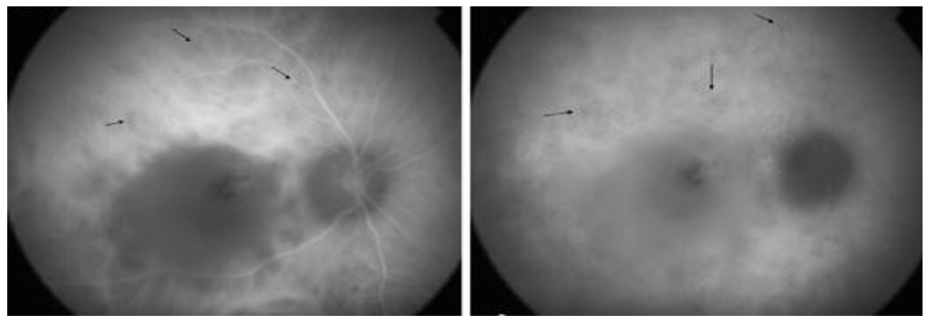
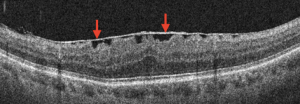
Age-related macular degeneration is a leading cause of severe vision loss worldwide in people over the age of 60. Although there is no direct link between exercise and macular degeneration, studies suggest that exercise can reduce the risk of developing the disease. This is because exercise can improve blood flow to the retina, which is essential for maintaining healthy vision.

In addition to reducing the risk of eye diseases, exercise can also improve energy levels, lower blood pressure and aid in weight loss. All these factors contribute to improved overall health and well-being. Regular exercise can promote a healthier lifestyle, leading to better rest for the body and improved quality of life.

In conclusion, exercise is beneficial not only for the body but also for the eyes. Regular exercise can significantly lower the risk of developing serious eye diseases. While exercise cannot single-handedly prevent or cure eye diseases, it can certainly help in your overall well-being. Additionally, it is crucial to get regular eye checks, as they can aid in the early detection of any eye disease. Don’t forget to visit our centre, OasisEye Specialists, where we provide comprehensive eye care services to help you maintain optimal eyesight.

Residents within Klang Valley in areas such as Cheras, Puchong, Shah Alam, Petaling Jaya, and Kepong can come visit our ophthalmologists to have their eyes examined at Nexus Bangsar South KL branch. Residents in Seremban 2, Senawang, Sendayan and Port Dickson can visit Dr Teh Wee Min at our Seremban branch. Residents in Johor Bahru, Skudai, Kulai, Iskandar Puteri, Senai, Tebrau, Batu Pahat, Kluang, Segamat can visit Dr Ling Kiet Phang or Dr Chan Choon Teng at our Johor Bahru branch. Residents in Perai, Bukit Mertajam, Butterworth, Penang island, Alor Setar, Kulim and Sungai Petani can visit Dato’ Dr Haslina at our Penang branch.

Men vs Women: Eye Health and Vision

It might come off astounding at how contrasting vision and eye health could be between men and women.
Take for example, men tend to succumb in sustaining sight-threatening eye injuries than women, however, women do tend to be more at risk to various eye diseases. What could both sexes do to address such eye concerns?

Common Eye Diseases in Women

Women are more susceptible to glaucoma and age-related macular degeneration (AMD) than men. The progressive loss of central vision is known as AMD, whereas, damage to the optic nerve would result in permanent vision loss known as glaucoma. Women are more susceptible to these diseases due to having longer life expectancies than men. Annual routine eye exams are beneficial for early detection hence, the best way to prevail them.
Besides the diseases mentioned above, women are also more prone to chronic dry eyes and basic refractive errors. When dry eyes are left as it is, it may lead up to an increased risk of eye infections. Symptoms of dry eyes include redness, discomfort, prickly sensations, irritation, and blurred vision.

Risk Factors for Eye Diseases

Age is a major risk factor in eye disease developments. However, for women this too includes pregnancy, birth control, as well as menopause. This is because it involves significant changes and fluctuations in hormone levels. Dry eyes is more anticipated due to these hormone changes too. Birth control may show indications of a woman’s chances in cataract development.

Eyes of Men and Hormones

In adolescence, pubescent boys may turn out nearsighted (myopic) for a short period of time as their eyes develop. As they undergo a growth spurt, their eyes may get longer. It should be noted that oftentimes this effect lasts into adulthood. Despite that, it can be simply resolved with spectacles or contact lenses. They may also consider refractive surgery such as TransPRK, ICL, etc. once they reach adulthood and their refraction stabilises.

Eye Injuries in Men

Comparatively to women, men are more susceptible to eye injuries as they are oftentimes more exposed to work in hazardous occupations or partake in high-risk sports. Men are also less inclined to wear protective eyewear and this facilitates the risk more when compared to women. Men too often tend to tough out their injuries and seek professional advice only after the injuries are intolerable. Whereas, women would have been more emotionally driven by the pain and anxiety to seek for advice sooner.

Vision in Men and Women

Both men and women actually see things differently. When it comes to distinguishing subtle differences in colour, women are better than men at it. Men in actuality are more commonly susceptible to colour blindness which are inherited from their mothers. In retrospect, men surpass women at identifying minute details and rapid movement. So don’t blame men too much if they can’t tell your lipstick shades apart!
One theory to account for these differences is that they go back to the hunter-gatherer days, Men did the hunting while women did the foraging, so the men would have needed to spot game at a distance in order to be effective hunters, whereas women needed to be able to tell nutritious plants apart from poisonous ones (especially green and yellow tones).

What Is Nystagmus?

Nystagmus is an eye condition where the eyes move rapidly and are unable to control. The areas of the brain that control eye movements are not working properly. There are three types of movement of nystagmus which are horizontal nystagmus (moving from side to side), vertical nystagmus (moving up and down), rotary nystagmus which move in a circular motion. This eye condition usually happens in both eyes and can occur in children or adults. The speed of movement may vary, it can be fast or slow and shake more in a certain direction. Therefore, you may have noticed nystagmus patients will tilt or turn their head to see clearer. This is because it helps to slow down the eye movements.

Types of nystagmus : Congenital and acquired.

Congenital nystagmus

Congenital nystagmus normally occurs in infants at the age of 6 weeks to 3 months old. It likely affects both eyes by moving horizontally (side by side). Congenital nystagmus causes are yet unknown, but sometimes it might be due to inheritance.

Acquired nystagmus

Acquired nystagmus usually occurs later in life. It can be caused by serious medical conditions, drugs or alcohol consumption.
Generally, children with nystagmus don’t feel their eyes shaking but notice some blur of vision. Unlike adults with nystagmus will often feel shaky when looking around.

Cause of nystagmus

In some cases the cause of nystagmus is still unknown. In other cases, nystagmus can be caused by other eye conditions.
Possible nystagmus causes :

  • Family history of nystagmus
  • Albinism
  • Certain eye disorders in infants or children (etc. congenital cataracts and binocular anomalies such as strabismus)
  • Multiple sclerosis
  • Inner ear problems (etc. Meniere’s disease)
  • Trauma or head injury (common happens in younger people)
  • Stroke (common happens in older people)
  • Alcohol or drug consumption
  • Use of certain medications (etc. lithium or anti-seizure medications)

Symptom of nystagmus

The main symptom of nystagmus is that eyes move rapidly and are unable to control. The movement usually is side to side. It also can be up and down or circular motion. The speed of movement can vary fast or slow and shake more in a certain direction.

In addition, other nystagmus symptoms may include:

  • Dizziness
  • Photophobia (sensitive to light)
  • Having difficulty seeing in dark environment
  • Blur of vision
  • Turned or tilted head position to see
  • Shaking environment

Nystagmus Treatment

Nystagmus are treated based on the underlying causes. Those who are born with nystagmus usually are not able to be cured. However, refractive correction with glasses or contact lenses is able to provide them a clearer vision which may help slow down the rapid movement.

Surgery repositioning the eye muscles that move the eyes is rarely done, and it does not correct or cure nystagmus. It only helps people with nystagmus to keep their head in a more comfortable position by limiting eye movement.

In acquired nystagmus sometimes it can go away by treating the underlying causes of nystagmus. For instance by treating the medical condition or stopping drug or alcohol intake.

Nystagmus can be diagnosed by an ophthalmologist. The ophthalmologist will examine the eyes to rule out any eye condition which relates to nystagmus conditions including binocular anomalies, cataract and any retina disease including optic nerve. Sometimes they also refer to an ear examination ,a neurological examination and brain imaging (computerised tomography (CT) and magnetic resonance imaging (MRI)) if necessary to rule out the underlying causes.

Therefore, it is advisable to visit an ophthalmologist as soon as possible if you notice any unusual sign or symptom of nystagmus especially for parents with infants or children. In addition, an eye screening can also be beneficial.

The impact of smoking on eyesight

Smoking your way to blindness

Tobacco smoking is the consumption of tobacco which can be found in lighting cigarettes, cigars and pipes. The practice of inhaling and exhaling such fumes poses an epidemic on its own, known most commonly to cause significant damage to the heart and lung. Ever thought how smoking could develop or exacerbate eye problems that are sight-threatening and potentially lead to blindness? Some of the eye diseases are such as:

Dry Eye Syndrome

Tobacco smoke irritates the conjunctival mucosa of the eye, decreases the quantity and quality of tears, thereby causing eye dryness. The insufficiency of quality tears causes smokers to experience itchiness, eye redness, foreign body sensation, swelling, and watery eyes.

Cataract

It is the clouding of the crystalline lens of the eye. Studies have found smokers significantly increase their risk of cataract development up to 3 folds as the practice of smoking causes alteration in the cells and increases build-up of heavy metal residue in the crystalline lens.

Age Related Macular Degeneration (AMD)

AMD is an acquired degenerative condition that impairs the central vision. Smoking causes constriction of the blood vessels and reduction in the level of lutein and zeaxanthin in the macula, which accelerates the rate of developing AMD by up to 4 times. It causes one to have difficulty in reading, seeing fine details, poor facial recognition, seeing wavy lines, and many more.

Uveitis

It is the inflammation of the central layer of the eye, known as the uvea. According to studies, smokers have an increased risk of 2.2 times to develop uveitis compared to non-smokers.

Glaucoma

Glaucoma is an eye disorder which is indicated by compromised blood flow to the optic nerve, resulting in progressive loss of nerve tissue and gradual vision loss. Smoking contributes to this condition by increasing plaque formation in the blood vessels, which in turn reduces blood flow.

Diabetes Retinopathy

It is established that smokers with Type 1 diabetes mellitus (DM) have a higher risk of developing diabetic retinopathy. The act of smoking combined with the chronic hyperglycemic state damages the tiny blood vessels in the retina by reducing retinal blood flow, causing these vessels to break, leak or even block.

Not only do smokers significantly increase their risk for all these diseases, it affects the eyes of people around them via passive exposure (secondhand smoke) as well. The negative impact of smoking during pregnancy can be transmitted to newborns, contributing to higher risk of visual disorders.

Residents within Klang Valley in areas such as Cheras, Puchong, Shah Alam, Petaling Jaya, and Kepong can visit our low vision specialist, Ms Vinodhini Naidu to have their eyes examined at Nexus Bangsar South KL branch. Residents in Seremban 2, Senawang, Sendayan and Port Dickson can visit Dr Teh Wee Min at our Seremban branch. Residents in Johor Bahru, Skudai, Kulai, Iskandar Puteri, Senai, Tebrau, Batu Pahat, Kluang, Segamat can visit Dr Ling Kiet Phang or Dr Chan Choon Teng at our Johor Bahru branch. Residents in Perai, Bukit Mertajam, Butterworth, Penang island, Alor Setar, Kulim and Sungai Petani can visit Dato’ Dr Haslina at our Penang branch.

What is Ocular Toxoplasmosis?

Toxoplasmosis is an infection caused by the intracellular protozoan parasite known as Toxoplasma gondii. The infection may be acquired or congenital, whereby the foetus acquires it from the mother while in the womb. Most people contract toxoplasmosis by eating raw or undercooked meat, vegetables or dairy products, or by coming into contact with infected cats, contaminated sandboxes or cat litter boxes – all of which have been contaminated with cat faeces. Although most parasite-infected individuals do not exhibit any symptoms, some people may experience flu-like symptoms, such as fever, headache and muscle aches. Infants and those with low immune systems are most frequently affected by serious forms of this disease. Toxoplasmosis during pregnancy may cause miscarriage and birth abnormalities.

Ocular toxoplasmosis occurs in the eye, where Toxoplasma gondii can cause inflammation. Frequently, ocular toxoplasmosis results from the reactivation of a congenital infection, although an acquired infection is now regarded to be more common. The parasite typically affects the retina causing eye pain, blurred vision and possibly permanent damage, including blindness.

What investigations are involved?

Ocular toxoplasmosis can look like other eye diseases that cause inflammation in the back of the eye (posterior and panuveitis). The doctor will look at the signs and symptoms to aid in coming to a diagnosis, which is based on clinical findings. If it’s not clear, a test called polymerase chain reaction (PCR) can be done to look for Toxoplasma DNA in the fluid from the eye and blood samples.
There are two imaging tests called Fundus Fluorescein Angiography (FFA) and Indocyanine Green (ICG) that can be used to visualize blood flow in the eye, which may help diagnose ocular toxoplasmosis.

Management / Treatment

Ocular toxoplasmosis requires personalized care, meaning that treatment will vary from person to person depending on factors such as the location of the infection, the severity of inflammation, and the patient’s immune system. If someone with a healthy immune system gets an ocular infection, it will usually go away on its own. However, if the infection affects certain parts of the eye, treatment may be necessary. People with weakened immune systems, such as those who have had transplants or are HIV-positive, may need long-term treatment.
There are several drugs available to treat ocular toxoplasmosis, and doctors often use a combination of drugs to help patients recover quickly and with minimal damage to their eyes. These drugs can kill the parasite responsible for the infection, but they work best when used together. In some cases, surgery may be necessary to treat complications like retinal detachment, cataract and choroidal or epiretinal neovascular membranes involving the macula.

It’s common for ocular toxoplasmosis to recur after someone has had it before, but relapses are not contagious and do not pose a risk to others. Relapses don’t pose a threat to an unborn child if the mother is pregnant.

Prevention

Preventive measures include thoroughly cleaning and washing fruits and vegetables, cooking meat adequately to destroy any harbored cysts, and avoiding contact with cat litter pans during pregnancy. Women of childbearing age must take adequate contraceptive measures for six months following primary toxoplasmosis infection.

In conclusion, preventing toxoplasmosis is crucial for maintaining healthy eyes, and early diagnosis and treatment are essential for managing the infection. At OasisEye Specialists, we offer comprehensive eye care services, including diagnosis and treatment of uveitis caused by toxoplasmosis. Our experienced team of eye specialists is committed to providing personalized care and innovative treatments to preserve and improve our patients’ vision. Contact us today to schedule a consultation and take the first step towards optimal eye health.

Contact Lenses

A contact lens is a transparent piece of “plastic” that sits on the front surface of the eye. It can provide access to clear vision, in addition to having various therapeutic usages. Like the idea of Cinderella being fitted with the perfect pair of glass slippers, finding the right pair of contact lenses is essential to ensure comfort throughout the duration of wear. How do we address this?

Before purchasing a pair of contact lenses, one should get their eyes checked with an eyecare professional, such as an ophthalmologist, optometrist or an optician with a Contact Lens licence. A thorough eye examination, which includes understanding your lifestyle, purpose of wear, vision and prescription check, and suitability of wear, is crucial to evaluate your suitability of wearing contact lenses. Contact lenses are great for improving your cosmetic appearance, flexibility of being glasses-free and useful for sports activities in which glasses are a hindrance.

Contact lenses are not a one-size-fits-all. The ideal best fit needs to be assessed for optimal fit and comfort. When a contact lens is fitted loosely, the lens might drop out or you may feel immediate discomfort upon insertion. Conversely, a tight contact lens may feel comfortable initially but become uncomfortable and cause eye redness after a few hours.

Contact lenses are available in a variety of lens wearing schedules, also known as modality. It refers to how long the contact lens can be worn after it is removed from its packaging and how soon it should be disposed or replaced. Replacement schedules could be daily, biweekly, monthly, 3 monthly, 6 monthly or yearly. As an example, daily disposable contact lenses should be discarded after a single day of wear and would be great for occasional wearers and perfect for a day out.

Do you know that there are various contact lens materials in the market? Depending on the material, the contact lenses may be soft, rigid or a hybrid of both. Examples of materials are hydrogel, silicone hydrogel, rigid gas permeable and many more. Each material has its own pros and cons, and therefore should be chosen based on your purpose of wear, desired duration of wear, refraction and ocular condition.

Retinitis Pigmentosa

What is Retinitis Pigmentosa?

Retinitis Pigmentosa, also known as RP, is a group of eye conditions that affect the retina. This condition can alter how the retina responds to light, making it difficult for people with RP to see. Essentially, RP causes changes to the cells in the retina known as photoreceptors, which do not work properly and can result in poor vision. The photoreceptors contain rods and cones cells, with rods being affected first in RP patients, causing difficulty seeing at night and loss of peripheral vision. RP causes a slow loss of vision over time, although most people do not become completely blind. RP usually affects both eyes and is often inherited from parents to their children.

Symptoms of Retinitis Pigmentosa

One of the most common early symptoms that retinitis pigmentosa patients experience is night blindness, which makes it difficult to adjust to darkness and see in low light conditions. Patients may also bump into objects in the dark. As the disease progresses, patients gradually lose their peripheral visual field, making it difficult to see objects to the side. In some cases, patients may also experience a loss of central vision in the later stages of the disease.

What Causes Retinitis Pigmentosa?

In most cases, Retinitis Pigmentosa is caused by changes in genes that control the cells in the retina. RP is usually due to genetic inheritance and can be linked to many genes in our body. It can also occur as part of other genetic conditions, such as Usher syndrome, which causes both vision and hearing loss and can lead to difficulty in balancing.

How is Retinitis Pigmentosa diagnosed?

A comprehensive dilated eye examination is required in order to diagnose Retinitis Pigmentosa. Clinical tests such as visual field, electroretinography (ERG), optical coherence tomography (OCT), and fundus autofluorescence imaging (AF) can also be used to diagnose RP. Genetic testing can also be done to determine the type of RP a patient has, which is important for family planning since RP is a genetic inherited disorder.

Treatment for Retinitis Pigmentosa

Unfortunately, there is currently no cure for Retinitis Pigmentosa. However, low vision aids and visual rehabilitation programs provided by low vision specialists can help RP patients improve their quality of life. Researchers are studying gene and cell therapies as potential future treatments for RP.

If you are experiencing any symptoms related to Retinitis Pigmentosa, we recommend visiting a retinal specialist at our centre. A retinal specialist will be able to determine if any diagnostic tests are necessary to rule out any abnormalities. Additionally, we offer genetic testing at our centre.

Epiretinal Membrane (ERM)

An epiretinal membrane (ERM) is an eye condition where a thin, clear, and transparent fibrous cellular material forms on the surface of the retina. It commonly occurs, affecting the posterior pole of the retina over the macula. The cause is unknown, but some research shows that it can be secondary to trauma, post-intraocular surgery, chronic ocular diseases, and so on.

Symptoms:

ERMs usually cause several symptoms, such as:

  • Difficulty in seeing fine details and recognizing faces when the central part of the retina is affected.
  • Blurred and distorted vision.
  • Straight lines appear wavy.
  • Decreased vision and loss of central vision.
  • Double vision.

Causes:

Epiretinal membranes can occur as a result of the normal aging process in the eyes. It is common in individuals over the age of 50. One reason for this condition is when the vitreous gel peels away from the retina. ERM may also form following retinal or eye surgery.

Diagnosis:

An epiretinal membrane can be diagnosed during a routine eye examination for mild cases where the symptoms do not affect the individual. During the eye examination, an optometrist or ophthalmologist may use a non-invasive imaging technique called Optical Coherence Tomography (OCT) to visualize the layers of the retina. In some patients, an ophthalmologist will request additional tests such as fluorescein angiography to determine any other underlying problem that might have caused this problem. The test involves the use of dye to light up areas in the retina.

Treatment:

In many cases, mild membranes are monitored over time for progression. Aside from surgery, there are no other available treatments for epiretinal membranes. Eyeglasses, contact lenses, and prescription eye drops are generally not effective treatments for epiretinal membrane.

Surgery is considered and recommended when the patient’s vision or perception of visual distortion starts to affect their quality of life, for example, when the vision is worse than 20/40 (6/12). To preserve the anatomic integrity of the retina, the membrane removal must be executed with extreme caution. The surgeon will make a tiny cut around 4 mm and remove the fluid from inside the eye. Then, the surgeon will hold and gently peel the epiretinal membrane from the retina and replace the fluid in the eye. The surgery is performed under local anesthesia.

Therefore, it is advisable to visit an ophthalmologist for a detailed eye examination if you experience any unusual symptoms related to an epiretinal membrane (ERM). Moreover, an eye screening can also be beneficial for the early detection of any retinal disorder.

Prosthetic Eye

An ocularist’s main duties are to create a prosthetic by moulding, colour matching and supplying the prosthetic to enable the patient some form of symmetry to the facial area. The aim of an ocularist is to replace the affected area cosmetically with the best symmetry possible. Ocularist try to create comfort for the patient, with as much movement as they possibly can of the prosthesis.

Eye plays a very important role in facial expressions. Ocular prosthesis rehabilitation can produce a significant impact for an individual’s physical, physiological, social and psychosomatic health for those who have a missing eyeball or loss of any tissue or organ. The reason for losing an eye can be due to the result of congenital defect, disorder, accidental trauma, serious ocular diseases, or surgical intervention. Surgical procedures for the removal of the eye can be classified as evisceration and enucleation. After the surgical procedure, the following treatment includes implant retained or a custom made acrylic ocular prosthesis.

A prosthetic eye can help to improve the appearance of the individual who has lost an eye. Wearing an eye patch or bandage is also one of the choices for individuals yet if the entire eye is removed, ocular implant and prosthesis prevent the eye socket’s tissues from growing into the empty space. A custom made ocular prosthesis is preferred as it provides close adaptation to the tissue surfaces and provides maximum comfort.

Prosthetic eye is commonly called or known as “glass eye” or “fake eye”. It acts as a shell that covers the structures of the eye socket. It does not restore vision so the vision will still have no perception of light (NLP).

What Is a Prosthetic Eye Made of?

“What is a prosthetic eye made of?” is a frequently asked question. A prosthetic eye can be made up of a few materials that can adapt with the eye socket tissue closely, such as hard, plastic acrylic. The common material used is acrylic plastic polymer which is also known as polymethyl methacrylate (PMMA). PMMA is lightweight, shatter-resistant and is an alternative to glass where glass was used at the beginning of this technology. Silicone polymers also used to make artificial eyes.

How is an ocular prosthesis created?

Prosthetic eye can be created when the eye socket is ready and stable after the surgery, approximately 3-6 weeks after the surgery. Prosthetic eye must be created by an ocularist who specialises in custom-crafting artificial eyes. There is a layer of the eye known as sclera, which is seen as the white of the eye. During the evisceration surgery, the damaged eye’s internal organs are removed while leaving the sclera intact. Inside the sclera is then placed an implant where it adds volume to the bony socket. Once the implant is put in place, the other layer of the eye is stitched. The eyelid needs to be closed together then a stitch is put in place for a week for 10 days. After 3-6 weeks of the surgery, the process of placing an artificial eye begins. The ocularist will measure the amount of space in between the eyelids and create a prosthetic shell. This is then hand painted such as it matches the colour of the normal eyes.

Adjustments and refitting of the prosthetic eye is needed as time goes on due to the fact that the eye socket can change in shape over time. Prosthetic eyes require yearly polishing by an ocularist and replacement every 5 to 7 years.

Low Vision Aids (Near)

Low vision aids are designed to improve visual performance and provide enrichment of daily experiences. Just like distance low vision aids, near low vision aids can be optical, non-optical and electronic devices.

Usually children tend to not complain about having difficulty with their near tasks due to their strong accommodation. However, when school activities increase and get more sophisticated with smaller reading material, greater magnification is required and objects need to be brought closer, making reading more fatiguing. Near aids can be beneficial when this becomes an issue for them.

Near Low Vision Aids:

Magnifying Spectacles

Advantages
– Hands free
– Larger field-of-view
– Prolonged reading can be done
– Binocular or Monocular depending on the individual’s need
– Portable
– Cosmetic appearance

Disadvantages
– Higher power, higher working distance
– Illumination will be obstructed (going closer will block the illumination)
– Close reading distance causes fatigue and uncomfortable posture
– Not effective for individuals with constricted visual fields
– Individual with eccentric fixation are unable to fixate through the spectacles

Hand magnifier with or without illumination

Advantages
– Convenient for short-term tasks
– Can use at comfortable reading distance
– Individuals with constricted visual field can benefit
– Available in both illumination and non-illumination

Disadvantages
– User unable to read and write simultaneously
– Maintaining focus is difficult
– Individual with tremors may not be able to hold it properly
– Visual field is limited

Stand Magnifier

Advantages
– Suitable for prolonged reading
– Beneficial for individual with shaky hands (tremors, arthritis)
– Available in both illumination and non-illumination.

Disadvantages
– Field of view reduced
– Bulky
– Posture affected
– Difficult to use if the surface is not flat

Solid Lenses Magnifier

Advantages
– Prolonged task can be done

Disadvantages
– Only available in low magnification
– Heavy

Visual Field Enhancement Devices:

Eccentric viewing Technique

A technique which can greatly improve a patient’s ability to determine objects and faces when a person views it peripherally with another area of visual field.

Non-Optical Devices

Typoscope – used as a guide for reading and writing for a person with a large visual field defect.

Extra Illumination – Different types of illumination needed varies for each individual and depends on their diagnosis and extent of pathology. High illumination required for most of the low vision patients. Direct lighting is useful for most of the patients and usually is advisable to place it 45 degrees with the visual axis for the better seeing eye.

Increase image size and print

Talking Devices

Electronic devices – Include closed-circuit televisions (CCTV), video magnifier systems, large print computer programmer and screen reader software such as JAWS and MAGic. With the advancement of technologies, electronic devices could benefit moderate to severe visual impaired individuals which normal optical aids or non-optical aids do not benefit them. The only drawback of electronic devices was most of the devices were expensive.

Low vision patients should undergo low vision rehabilitation for proper training, environmental modification, orientation, mobility training, counselling and selection of suitable low vision aids to accomplish vocational and avocational activities independently. Visual aids help to improve both sight and quality of life for low vision patients.