Low Vision Aids (Distance)

Low vision aids are designed to improve visual performance and provide enrichment of daily experiences. Low vision aids can be optical, non-optical and electronic devices.

Distance Optical Aids:

When conventional lenses cannot provide required visual range, aids that have optical properties will be capable of promoting better visual performance. Telescope is an optical instrument that improves the resolution of an object by increasing the size of the image projected on the retina by making things at distance appear to be closer. The use of telescopes can help individuals in their daily and social activities such as watching television, reading white boards, street signs, finding entrances from a building bus number, traffic signal, building numbers, billboards and menu boards. Advantages of the telescope as low vision aids include being simple and easy to bring along while the disadvantages are restriction of visual field, illumination, difficulty in locating and focusing on objects quickly, only suitable for short term viewing, cannot be used when mobile, limited depth of focus and expensive. In order to use telescopes more effectively, special skills and training are required.

Types of telescope

  • Keplerian or Galilean
  • Monocular or binocular
  • Hand-held, spectacle-mounted or clip-on

Keplerian Telescope

An optical system that uses two convex lenses. Image produced by Keplerian was real and inverted which required a prism to reverse the image. Therefore it is heavier, longer and costs more compared with Galilean. Keplerian provides a greater visual field and better optical quality than Galilean.

Galilean Telescope

An optical system that uses one convex lens which is closest to the object and one minus lens which is closest to the eye. Image produced by Galilean is real and upright. It is lighter, shorter, and cheaper than the Keplerian. Thus, is the first-choice prescription. It is suitable for people with peripheral field loss due to it providing a wider visual field.

Monocular or Binocular

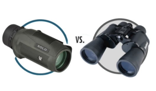
Monocular telescope is indicated when there is a significant difference in visual acuity between the 2 eyes. It is advisable to use in the better-seeing eye. It is more compact, lightweight, and cheaper. Drawback associated with the monocular telescope was eye fatigue and lesser field of view compared to the binocular telescope. Binocular telescope is indicated when there is similar visual acuity in both eyes, with the purpose of increasing the visual field, and for nystagmus. Binocular telescopes will be heavier and expensive.
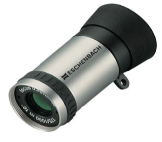
Hand-held telescope

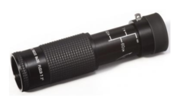
It is light, compact and cheaper than the Galilean and Keplerian. It is suitable for short activities and could be a first prescription choice.


Spectacle-mounted telescope

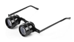
It is suitable for those who have hand-eye coordination problems and is very useful for prolonged activities or activities that require visualisation of details.

Clip-on

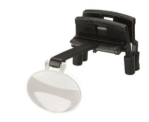
The clip-on model has both advantages. It is lighter than the spectacle-mounted model. However, the lenses can be scratched and the visual field can be reduced to further distances.
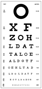


Effect of Alcohol to the Eyes

Alcohol, a psychoactive substance with dependence-producing properties, has been widely used in many cultures for centuries, according to the World Health Organization (WHO). Although alcohol consumption is often associated with people having a good time and enjoying themselves, taking it too far can have serious consequences on your health.

Most people associate heavy drinking with liver disease or accidents, but did you know that alcohol can also have an impact on your eyes? Excessive alcohol consumption can cause both short and long-term effects on your vision and eye health, and it’s important to be aware of both.

Short-term effects of alcohol on your vision

Short-term effects of alcohol on your vision can impair your peripheral vision and lead to tunnel vision. Alcohol hinders your pupils’ ability to react, preventing them from constricting or dilating properly. Car headlights, for example, can directly slow your reaction time, making it challenging to do simple tasks like driving at night. This is one of the key reasons why it is risky and against the law to drive after drinking.

Furthermore, alcohol can cause blurry and distorted vision, which everyone reacts to differently depending on the amount consumed. There is a delay between the brain and the eyes due to the slow rate of communication between neurotransmitters in the brain, which weakens eye muscle coordination. Alcohol can also affect one’s capacity to perceive contrast, reducing it by 30% at the permissible blood alcohol level, according to a study. Contrast is the visual system’s processing of differences in brightness and is the root cause of this temporary impairment, making it more difficult to distinguish between different objects based on lightness and darkness, such as traffic lights.

Excessive or regular alcohol intake can also cause dry eyes and myokymia, or twitching of the eyelids, resulting in temporary inflammation, double vision, eye burning and itching, migraines, and sensitivity to light. The long-term consequences of these symptoms include expanded blood vessels in the eyes, making them often appear red and bloodshot.

Excessive or regular alcohol intake can also cause dry eyes and myokymia, or twitching of the eyelids, resulting in temporary inflammation, double vision, eye burning and itching, migraines, and sensitivity to light. The long-term consequences of these symptoms include expanded blood vessels in the eyes, making them often appear red and bloodshot.

Long-term effects of alcohol on your vision

Regular alcohol consumption over time can also increase the risk of early cataract formation, which can start to manifest as early as age 40. Long-term impairments may also include permanent blurring of vision or double vision caused by the weakening of the eye muscles, resulting in a slower reaction time.

Moreover, prolonged alcohol abuse can affect your vision due to vitamin deficiencies. Heavy drinking can interfere with the liver’s ability to absorb vitamins essential for maintaining healthy eyes and good vision. Alcohol can only be broken down by the liver so quickly.

Drinking too much alcohol may make you more susceptible to age-related macular degeneration (AMD). According to WHO research, the average person consumes 33g of pure alcohol every day, which is equivalent to two glasses of wine. Based on an Australian study, people who drink more than 20g of alcohol per day have a 20% higher risk of developing early AMD than those who don’t drink at all.

Optic neuropathy or optic atrophy is one of the most dangerous effects of long-term alcohol use on vision. This disorder, brought on by excessive drinking or smoking, is also known as tobacco-alcohol amblyopia or toxic amblyopia. It causes a painless loss of eyesight, as well as diminished colour vision and peripheral vision. Alcohol increases the risk of heart disease, and heart disease symptoms can be seen in the eyes, including optic neuropathy, atrophy, bleeding in the retina from vascular occlusions, and even hypertensive retinopathy.

What causes bloodshot eyes?

What does the term bloodshot mean? It basically means the blood vessels under the eye’s surface become larger and inflamed, causing the eye to look red. Some red eyes alone are usually benign and have no reason for concern. But if the redness is accompanied with pain or loss of vision, this may indicate a serious medical problem and may need immediate medical intervention.

Listed below are some of the causes for eye redness:

Allergies

One of the most common causes of redness is allergens in the surroundings. Outdoor allergens such as pollen from plants and trees. Indoor allergens consist of dust, pet fur, pet dander and even mold. The redness is often paired with itchiness, sneezing and teary eyes. This is referred to as allergic conjunctivitis, which isn’t contagious.

Dry eye

When the lacrimal glands do not produce enough tears to lubricate the eye, this can result in dry eyes. Sometimes some hormonal medications (HRT) such as estrogen can cause dry eyes as a side effect.

Subconjunctival Haemorrhage

Our conjunctiva contains many capillaries and blood vessels. If these vessels break, blood can leak into the white and the conjunctiva, which then builds up in a clump to cause subconjunctival haemorrhage. Sometimes, the simple action of coughing or sneezing can cause this to happen. People who have medical conditions such as diabetes, high blood pressure and are on blood thinners are more prone to this occurring. These bleeds may look serious, but they are usually painless and do not affect vision. It will resolve within 7-10 days.

Conjunctivitis (Pink Eye)

Conjunctivitis is the infection of the thin transparent membrane of the eye called conjunctiva. The most common ones are viral and bacterial conjunctivitis. Both are equally contagious and can spread easily. These are sometimes accompanied by a sticky discharge, watery eyes, a gritty burning sensation, sensitive to bright light or crusting of the eyelids in the morning.

Blepharitis

This is when your eyelids are inflamed, which causes your eyelids/eyes to be swollen and red. The symptoms may be similar to those of conjunctivitis, so it is advisable to get it examined by a doctor to determine what is the cause of your red eye.

Alcohol

Excessive drinking of alcohol can lead to bloodshot eyes. The alcohol content causes vasodilation of blood vessels, so more blood gets filled into these dilated vessels, thus making the eye look red. Alcohol is also a type of diuretic which makes the body (and the eye) dehydrated after frequently urinating. When the eye is dehydrated, it’ll then lead to dry eyes.

Contact Lens Wear / Corneal Ulcers

People who wear contact lenses touch their eyes more often than those who do not. Thus, they are more prone to getting eye redness, if not, eye infections. Sometimes, removing contact lenses can result in scraping or scratching the cornea. Overwearing of CLs over the given timeframe may lead to neovascularization (redness). Sleeping with CLs, or unhygienic practice of cleaning CLs may lead to corneal ulcers. Action must be taken immediately if corneal ulcers arise.

Acute angle closure glaucoma

There are different types of glaucoma, but one to take note of in regards to eye redness is acute angle closure glaucoma. The redness is usually accompanied by an intense pain in the eye, haloes/rainbow rings around light, blurred decreased vision, headaches and nausea/vomiting. The iris of the eye prevents eye fluid from being drained out properly, and as the fluid builds up, this will increase eye pressure rapidly. This is usually an emergency case and has to be seen by a doctor immediately to help to bring the eye pressure down to prevent vision loss.

Episcleritis/Scleritis

Episcleritis is the inflammation of the layer between the conjunctivitis and sclera. Symptoms consist of mild pain, redness and tenderness when touched. This is easily treated with artificial tears, and if needed, steroids to keep the symptoms at bay. It will usually resolve within 10 days.
On the other hand, Scleritis is the inflammation of the sclera which causes the eye to be very red. This is usually caused by an autoimmune condition, so it will need immediate medical attention. Patients will be given anti-inflammatory medication and topical steroid drops.

Uveitis

This is the inflammation of the middle layer of the eye called the uvea. Symptoms come in the form of pain, redness, blurred vision, and sensitivity to light. This must be treated immediately with steroids to bring the inflammation down. If not treated, it may cause retinal/choroidal scarring or even uveitic glaucoma.

Injury – trauma to the eye

Simple actions like accidentally poking your eye with your mascara applicator, or kids playing roughly and fingers poking each other’s eyes, can result in injuries to the eye. When the eye is injured, blood vessels in the eye will dilate to bring more blood cells to the site of injury to repair and heal the injury.

Swimming

Spending a certain amount of time in the pool can cause bloodshot eyes as there is chlorine in the pool which then irritates the eyes.

Lack of sleep

Not getting enough sleep can also lead to red eyes. Not enough rest means no proper blood/fluid circulation, so this makes your eyes look puffy and red.

Smoking

Cigarettes contain harmful toxic chemicals which are irritable for the eyes. When the eyes get in contact with these chemicals they will react and cause their eyes to be red.

All in all, as long as you have a red eye (which sometimes may be accompanied with pain, discharge or blurred vision), it is advisable to seek medical attention to determine the cause and treat it when necessary, to prevent other issues from arising.

Swimming with Contact Lenses

Swimming with contact lenses on may seem like a great idea because without them, you cannot truly see clearly while you’re out splashing away in the waters. However, by doing so you risk numerous eye issues which could be dangerous and may not be worth it after a fun day at a pool or beach.

Here are insights regarding the risks of swimming with contact lenses on and precautions you can take to protect your eyes better in the water.

What are the Risks Swimming with Contact Lenses on?

The FDA does not advise for contact lenses to be exposed to any kind of water. This is due to the nature of soft contact lenses to absorb water, trapping potential bacteria, viruses, or other pathogens on the eye which makes the wearer susceptible to certain eye issues when worn to swim at the pool or beach.

On another note, swimming in a pool may be less eye threatening than swimming in lakes, rivers and out at sea. The reason may appear obvious as water in lakes, rivers and the sea contain a variety of bacteria, viruses, and other harmful pathogens that often would be mostly eliminated by pool chemicals.

However, that does not mean wearing contact lenses to swim at the pool is safe. In fact, chlorine and other pool chemicals are not able to completely eliminate pathogens. Rigid gas permeable (RGP) contact lenses should never be worn to swim, as they are easier to dislodge from your eye. Soft contact lenses on the other hand are more likely to remain on your eye when swimming, but they are porous and can absorb chemicals and bacteria, increasing the risk of eye irritation and infection.

Wearing your contact lenses while swimming could increase your risk of the following issues:

  • eye irritation due to lens adherence on the eye
  • dry eyes, especially after swimming in chlorinated or salt water
  • eye infections
  • corneal abrasion (scratched cornea)
  • eye inflammation (uveitis)
  • corneal ulcers

What about Showering with Contact Lenses On?

The risk of this is similar to swimming with contact lenses on. According to the Centre for Disease Control and Prevention (CDC), there’s a type of microorganism called Acanthamoeba which is found in all types of water. It is more commonly found in tap water and well water, which are the ones you shower with.

This amoeba may cause a serious eye infection called Acanthamoeba Keratitis which presents as eye pain and can be very difficult to treat. In rare cases, it may even lead to blindness.

Tips for Swimming Safely

  1. Goggles
    Swimming goggles work well enough to reduce the risk of eye infection if you are going to wear contact lenses anyway. Not only does it protect your eyes from contaminants, swim goggles also helps to prevent your lenses from coming off.However, it is still highly recommended to take your contact lenses out before coming into contact with water.

    In the case where your vision is poor without corrective lenses, you could consider investing in a pair of prescription swimming goggles. These could too come with UV protection which would be an added benefit against sun damage to the eyes.

  2. Refractive Surgery and Other Alternatives
    Many of those, typically with an active lifestyle and sporty, choose to undergo refractive surgery to correct their myopia (shortsightedness), hyperopia (longsightedness), or astigmatism with laser eye surgery. With this, you can say goodbye to glasses and contact lenses.There are various types of laser refractive surgery such as; TransPRK, LASEK, LASIK and various other technologies. Each to their own reshapes the cornea using a computer-controlled laser beam. Refractive surgery allows light to enter the eye and properly focus on the retina for clear vision. These surgeries have a good safety profile with most people achieving 6/6 vision without glasses or contacts after the laser procedure. Nevertheless, all surgeries come with possible risks and complications, which needs careful consideration before undergoing the procedure.

    Another alternative is Ortho-K, or orthokeratology. It is a non-surgical method to temporarily correct your refractive error and reduce the need to rely on glasses or contact lenses. Generally it is a type of hard contact lenses that are specifically designed and fitted to reshape the cornea. Typically worn at night while you are asleep, it allows you see clearly when you remove the lens in the morning when you’re awake. It is a safer non-permanent alternative for those who are concerned about permanent refractive surgery.

Your eye doctor or eye care practitioner would be able to better advise you on alternatives to swimming with eye correction to accommodate your lifestyle.

Hydroxychloroquine Drug Toxicity to Eye

Hydroxychloroquine is a controlled medication for several dermatologic and rheumatologic conditions. It is also widely used to treat Systemic Lupus Erythematosus (SLE) disease and Malaria (by using Chloroquine originally). The maximum recommended dose is 5 mg/kg/day which is commonly used in SLE patients. Recent research trials suggested that high dose HCQ is a possible potential use for coronavirus, COVID-19 therapy by reducing viral replication, but there is insufficient evidence supporting its efficacy. Ongoing long term usage and high dose of hydroxychloroquine can cause unwanted side effects to the body including eyes.

Side effects

  • Dizziness
  • Headache
  • Ocular toxicity
  • Skin rash
  • Gastrointestinal upset such as diarrhoea, vomiting and stomach cramps

In severe cases seizures, angioedema, arrhythmia, and bronchospasm may occur. Long term hydroxychloroquine usage can cause toxicity to the eyes as well including the cornea, ciliary body and retina.

Eye diseases

Chloroquine was initially used to help treat malaria. Chloroquine is less often used now because its derivative hydroxychloroquine is better. Hydroxychloroquine is commonly used to treat inflammation in diseases like rheumatoid arthritis and dermatitis. The toxic effects of this substance on the retina are seen in the area known as the macula. Some people may have early signs of toxicity, such as changes in their vision. More advanced toxicity may involve complaints like difficulty seeing in colour or a scotoma (a dark spot on the centre of your vision). Advanced hydroxychloroquine toxicity can cause a bull’s eye maculopathy. Since retinal toxicity is usually irreversible, finding out if someone has retinal toxicity and stopping the thing that is causing the toxicity as soon as possible is the best treatment. Corneal toxicity is when a drug gets deposited into the cornea. This is very rare, and it typically doesn’t affect vision. In rare cases, ciliary body dysfunction can happen and influence the eye from focusing.

Risk factors

Hydroxychloroquine retinopathy is more common in patients with longer duration and daily dosage of the drug. Patients are at higher risk of retinopathy if they use hydroxychloroquine for more than 5 years as treatment.

Signs

Hydroxychloroquine retinopathy is usually bilateral and symmetric. The early signs include macular edema and granular depigmentation in the retinal pigment epithelium in the retina. It can progress to an atrophic bull’s eye maculopathy if continuous drug exposure causes widespread other areas of the retina including optic nerve. Hydroxychloroquine keratopathy is seen as an intraepithelial deposit. Ciliary body dysfunction can be detected when the near vision is poor.

Symptoms

Initially, hydroxychloroquine toxicity may cause few or no symptoms. However, over time, symptoms may develop such as hard to differentiate colour, loss of central vision, reading difficulty, blur of vision, glare, flashing of lights, and metamorphopsia. It usually affects both eyes. In keratopathy, patients may experience halos around light and photophobia whereas in ciliary body dysfunction, will complain of reading difficulty and find it hard to focus on near work.

Treatment

Early detection of hydroxychloroquine toxicity can be stopped to prevent further retinal damage and visual loss. Patients who start hydroxychloroquine as medical condition treatment should be on regular eye examination, especially those who are on exposure drugs for more than 5 years. There is no treatment that can be done by surgery at the moment. Hydroxychloroquine retinopathy is not reversible as the damage appears to continue for a period of time. However, the early detection of retinopathy can prevent further visual deterioration. It can be treated in keratopathy cases. If you are a patient with ongoing hydroxychloroquine, it is advisable to have an eye examination with an ophthalmologist to rule out any eye disease.

What is Perfect Vision: 6/6 or 20/20?

People claim to have perfect eyesight if they have 20/20 vision or 6/6 vision. What does it actually mean?

The sole difference is the unit of measurement in feet and metres, respectively. When at 6 metres, and being able to see what a regular individual can see at 6 metres, the person would have 6/6 vision. Whereas, 20/20 vision depicts the same function, albeit at 20 feet instead of 6 metres. In Asia, these measurements are used interchangeably.

The visual system functions in the way that it processes visual information for us to be able to see features and images at far distances. We inherit the integrity that builds our visual system from our parents and the way it develops during early childhood. Within a populace of individuals with healthy eyes, the prime vision achieved would

vary from slightly better than most (6/4 or 6/5) to slightly poor (6.7.5). An example of these in our daily lives are; an individual is legally allowed to drive in Malaysia if they could attain a vision measurement of 6/12 in their better seeing eye with corrective spectacles.

What assessments can test for 6/6 or 20/20 vision?

The examiner will start the vision testing by asking you to read letters from the chart (commonly known as a Snellen chart). The test will be tested one eye at a time, and then with both eyes. The examiner can then measure your vision.

These results will provide important information regarding your vision and the prescription necessary to correct it.

Why is having good vision so important?

  • Safety: By having a poor vision, activities such driving, biking, cycling and even walking can be dangerous for you and others. In Malaysia, the minimum vision requirement is 6/12 (20/40) vision or better to be considered safe to drive on a public road.
  • Comfort: A clear vision allows you to have more freedom in your daily activities. Having good vision means that you won’t have to squint, whether you’re sitting at the back of the classroom or at the back row of the film theatre. You don’t have to strain your eyes to see things if you have good vision.
  • Reading ease: Having good vision enables you to read comfortably and learn better.
  • Quality of life: A good vision will provide you a better quality of life.

What causes you to have poorer than 6/6 or 20/20 vision?

Basically, there are various possibilities that can cause vision to be worse than the optimum 6/6 or 20/20 vision. The most common one are refractive errors such as:

  • Far-sightedness (hyperopia)
  • Near-sightedness (myopia)
  • Presbyopia: A condition that makes it more difficult to focus on a close up object as you get older, due to aging
  • Astigmatism: A condition caused by irregularity of the front part of the eye (cornea)

However, there are also many different eye disorders or diseases that can affect your eyesight, especially for those who are having systemic disease such as diabetes and hypertension.

How can your vision be improved if it is not 6/6 or 20/20?

There are several ways to help eyes to see clearly after you get your vision tested by an eye practitioner. The eye care professional with advice on the treatment options that fit and work the best for you, which may include:

  • Glasses: Glasses or spectacles are the most conventional way of vision correction. It is very practical, affordable and safe. They work by refracting the light to focus on the retina, which helps you to see clearly.
  • Contact lenses: Contact lenses work the same way as glasses to provide you a clear vision. However, it is more convenient and suitable for physical activities, especially for those who have an active lifestyle. Some people also prefer contact lenses for cosmetic purposes. Also, in certain refractive error conditions, contact lens correction can give better vision. Example, for those who have a large difference in refractive error or “power” between both eyes.
  • Refractive surgery: Refractive surgery is performed by changing the refractive, or light-bending, properties of the eye. It is functional because it allows you to see clearly without depending on glasses or contact lenses. However, some may not be good candidates for refractive surgery due to various reasons. Examples of refractive surgery include Implantable Collamer Lens (ICL), laser treatment such as TransPRK, and cataract surgery.

What will happen to your vision as you get older?

As you get older, your vision may be affected, despite having 6/6 or 20/20 vision at distance throughout your younger years. Those in their 40s will begin to have some difficulty focusing on close objects or small print, and some might have difficulty telling colours apart. Two common eye conditions caused by aging are presbyopia or cataract

When should you visit an eye practitioner or eye doctor?

You should visit an eye practitioner or eye doctor when you have a condition such as: ● Eye pain

  • Blurry vision
  • Headaches due to eye strain
  • Eye redness
  • Eye swelling

What is Low Vision?

Low vision is a condition where decrease in visual acuity or visual field constricted to the point where glasses, contact lens, medical or surgical treatments cannot be fully corrected. Low vision is a vision problem which will interfere with an individual’s daily activities such as driving, reading, etc. The National Eye Survey 1996 estimated that there were 540,000 people with low vision and about 50,000 people who were blind in Malaysia. Prevalence of low vision was found to be 2.44% and those who were blind were 0.3%.

What causes Low Vision?

It is often caused by eye diseases or conditions such as: cataract, age-related macular degeneration, diabetic retinopathy, glaucoma, retinitis pigmentosa, albinism, retinopathy of prematurity, amblyopia, strabismus, uncorrected high refractive error and eye injuries.

Symptoms & Types of Low Vision

  • Loss of central vision
  • Loss of peripheral vision
  • Blurred vision or hazy vision
  • Night blindness
  • Decrease in contrast sensitivity
  • Difficulty differentiating colors
  • Difficulty recognizing objects at a distance and near

Treatment

Unfortunately, low vision is usually permanent. An individual who was diagnosed with low vision can undergo low vision therapy. Low vision therapy will be done by an optometrist or an occupational therapist who specializes in low vision. The purpose of low vision therapy is to help individuals continue performing their daily activities. This is achieved by providing appropriate optical devices and special training in the use of residual vision and low vision aids, which range from simple optical magnifiers to high magnification video magnifiers. Low vision devices can be optical (telescope, magnifying glasses, magnifiers, etc.), non-optical (extra illumination, typo scope, books with enlarged text, writing guides, clock with large numbers, keyboard with large letters, etc.) and electronic device (Closed-circuit television (CCTV) or portable electronic devices). There are some resources centers that can help low vision patient such as:

  • Social Welfare Department (JKM)
  • Ministry of Women
  • Family and Community Development, Malaysia
  • Special Education Division, Ministry of Education, Malaysia
  • Malaysian Association for the Blind (MAB)
  • Society for the Blind Malaysia (SBM)
  • St. Nicholas Homes Center
  • Sabah Society for the Blind (SSB)
  • Sarawak Society for the Blind (SKSB)

Nutrients & Vitamins for Eye Health

Nutrition play an important role in slowing the progression and preventing the onset of ocular diseases such as Age-related Macular Degeneration (AMD), glaucoma, cataract and so on. Studies show getting sufficient essential nutrients can preserve good vision and eye health. Antioxidant-rich foods and supplements such as anthocyanins, carotenoids, flavonoids, and vitamins have been shown to lower the risk of getting eye-related diseases such as Age-related Macular Degeneration (ARMD).

  • Vitamin A
    • A study showed the reduction of glaucoma risk among a population of black women with intake of fruits and leafy green vegetables high in vitamin A. The sources of vitamin A include dark green produce such as kale, mustard, collard, and broccoli, orange produce such as carrots, sweet potatoes, papaya, egg yolks and whole milk.
  • Vitamin C
    • Vitamin C functions as an antioxidant and it can lower the risk of developing early cataract and helps to slow down the progression of age-related macular degeneration (AMD) and vision loss. The sources of vitamin C are citrus fruits, red and orange produce such as tomatoes, cantaloupes, peppers, green produce such as broccoli, cabbage. Other than that, cauliflower and berries are also rich in vitamin C.
  • Vitamin E
    • Vitamin E works well together with vitamin C to protect against the unstable molecules (free radicals) that damage the healthy tissue on your eyes. The sources of vitamin E can be found in egg yolks, avocados, butter shrimp and dark green leafy vegetables.
  • Lutein and Zeaxanthin
    • Many studies show that lutein and zeaxanthin lower the risk of age-related macular degeneration (AMD) and cataract. The sources of lutein and zeaxanthin can be found in dark green leafy vegetables and fruits such as broccoli, peas and corn.
  • Zinc
    • The human eye contains high levels of zinc which plays an important role in eye health. Zinc helps to transport vitamin A from liver to the retina to produce melanin where melanin acts as a protective pigment in the eye. The sources can be seafood, nuts, seeds, cheese, yoghurt, soybeans and so on.
  • Essential fatty acids
    • Essential fatty acids aid visual development and retinal function. It functions by fuelling cells and optimising central nervous system function. Omega-3 fatty acids, particularly eicosapentaenoic acid (EPA) and docosahexaenoic (DHA), are vital in vision development and maintaining proper eye and retinal function. Sources of omega-3 fatty acids include cold water fish such as salmon and sardines.

A sufficient amount of nutrients & vitamins play an important role for a healthy eye. However, excessive intake of vitamins or supplements may sometimes reverse the effect causing unnecessary problems as well. For example, high doses of beta carotene (Vitamin A) intake can lead to lung cancer. Therefore, it is encouraged for people who are having vision problems and with poor control of diet to seek advice from eye professionals for the right food to maintain their eye health. An eye health professional can help you create a diet plan which is suitable for you and helps to reduce the chances of developing age-related eye diseases.

Is Headache Related to the Eyes?

One of the most common symptoms that many people face is a headache. Even if you think you can treat a headache on your own without seeking medical attention, you should be concerned if the headache is accompanied by symptoms like blurred vision, slurred speech, weakness, numbness, or seizures. Genes can play a role in headaches, especially migraines.

Headaches can also affect the eyes and vice versa. That much is true. When you have a headache, the area around your eyes can occasionally hurt. Here are a few types of headache that are linked to the eyes.

  1. Eye strain headaches are triggered by poor posture, lack of sleep, being hungry or dehydrated or when your eye muscles contract too much when working for prolonged periods at near work. It will cause pain and discomfort behind your eyes, on one or both sides of your head, and from your neck down to your shoulders.
  2. Ocular migraines cause visual disturbances with or without headache. The cause is unknown, but genetics can be a contributing factor. People with ocular migraine may experience symptoms like seeing flashing lights, blind spots in the field of vision, severe eye pain, and headaches.
  3. Uncorrected refractive errors such as short-sightedness (myopia), long-sightedness (hyperopia), astigmatism, and presbyopia also interfere with proper vision and may cause squinting to help the eyes focus. If left untreated, it may also cause headaches.
  4. A sinus headache can develop due to a sinus infection. Many people don’t realise that having sinusitis means not only feeling pressure around the nose area. Actually, the sinus pressure also occurs around the eyes, cheeks, and mouth, which gives the same feeling as a headache.
  5. Headaches due to glaucoma most commonly occur around the eye or forehead. Glaucoma is divided into two types, which are primary open angle glaucoma (POAG) and acute angle closure glaucoma (AACG). One of the symptoms of AACG is headache.
  6. Ocular Ischemic syndrome develops due to a chronic lack of blood flow to the eye. People having this problem might have symptoms like headaches and a decrease in vision.

Home remedies for relieving headache symptoms:

  • Taking over-the-counter painkillers is able to relieve mild to moderate headaches. If you have a severe headache, do see a doctor immediately
  • Resting in a dark room
  • Apply cold compress to the back of the neck, the forehead or the eyes
  • Follow the 20:20:20 rule to relieve eye strain due to long-term near-work
  • Sitting with good posture
  • Drink more plain water

Despite the fact that headaches are relatively common, they should not be taken lightly as daily activities can be interrupted. To rule out any eye disorders, do see an ophthalmologist. The sooner you receive a diagnosis, the sooner you can start receiving treatment to relieve your symptoms.

Hypertensive Retinopathy

The retina is the clear photosensitive tissue that is located at the back of the eyes. It helps to process and transform light into nerve signals. The nerve signals will be interpreted in the brain, providing visions. Serious medical conditions could affect the retina, such as high blood pressure (hypertension).

Hypertensive retinopathy (HR) is an eye disease in the retina due to complications of high blood pressure (hypertension). Poor control of high blood pressure may cause vascular disease changes without being aware of it. As a result, the retina’s blood vessel walls will thicken, causing the blood vessels to become narrow and restrict the blood flow reaching the retina. Over time, high blood pressure can limit the functioning of the retina by putting pressure on the optic nerve. Eventually, it can cause vision problems. For this reason, it is essential for patients with high blood pressure to have a regular eye check.

Older people are at a higher risk of developing high blood pressure and, therefore, most likely to develop hypertensive retinopathy. The following conditions can also increase your risk of hypertensive retinopathy: They include prolonged high blood pressure, heart diseases, atherosclerosis, diabetes, smoking, high cholesterol, obesity, having an unhealthy diet, having a family history of high blood pressure, and heavy alcohol consumption.

Symptoms and signs

Generally, a person with mild hypertensive retinopathy might not experience any or very mild symptoms. The patient usually becomes more symptomatic as it progresses and in the late stages. However, an ophthalmologist can detect early stages by identifying vascular changes during fundus examination.

  • Common symptoms include:
  • Blurred vision
  • Subconjunctival haemorrhage (bleeding in the white part of the eye)
  • Double vision accompanied by a headache

Controlling high blood pressure is the only way to treat hypertensive retinopathy. Therefore, lifestyle changes that include quitting smoking, exercising regularly, losing weight, dietary changes, and reducing alcohol intake help to reduce the risk of this disease.

However, individuals with hypertensive retinopathy are at risk of developing various complications. This includes the following:-

  • Retinal artery occlusion: It occurs when blood clots block the arteries that carry blood to the retina. This results in insufficient oxygen or blood supply in the retina, which may cause loss of vision.
  • Retinal vein occlusion: This condition occurs when a blockage in the retinal veins prevents blood from being carried away from the retina. A blood clot in the veins causes it.
  • Ischemic optic neuropathy: This condition occurs when high blood pressure blocks normal blood flow within the eyes and leads to damage to the optic nerve, which responsibly transmits images to the brain.
  • Malignant hypertension: This condition occurs when a sudden increase in blood pressure interferes with vision, causing sudden vision loss. However, this condition rarely happens but it could be potentially life-threatening.

Hypertensive retinopathy is also associated with an increased risk of stroke and heart attack. Prevention of hypertensive retinopathy is possible by carefully managing high blood pressure and related conditions like diabetes. Lifestyle changes such as quitting smoking, losing weight, exercising regularly, and having a healthy diet could also help prevent hypertensive retinopathy. People with high blood pressure should monitor their blood pressure regularly and have an annual eye screening. It is urged to visit an ophthalmologist as soon as possible if there is a significant sign and symptom of hypertensive retinopathy.